Hot Topics and Position Statements
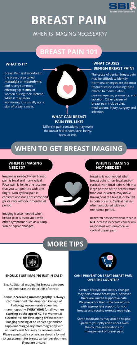
Abstract: Breast pain (or tenderness) is a common symptom, experienced by up to 80% of women at some point in their lives. Fortunately, it is rarely associated with breast cancer. However, breast pain remains a common cause of referral for diagnostic breast imaging evaluation. Appropriate workup depends on the nature and focality of the pain, as well as the age of the patient. Imaging evaluation is usually not indicated if the pain is cyclic or nonfocal. For focal, noncyclic pain, imaging may be appropriate, mainly for reassurance and to identify treatable causes. Ultrasound can be the initial examination used to evaluate women under 30 with focal, noncyclic breast pain; for women 30 and older, diagnostic mammography, digital breast tomosynthesis, and ultrasound may all serve as appropriate initial examinations. However, even in the setting of focal, noncyclic pain, cancer as an etiology is rare.
https://doi.org/10.1016/j.jacr.2017.01.028
Patient and Provider Resources

Patients often have questions about breast cancer screening, so we’ve created a handout with answers to frequently asked questions. You can access the provider handout here and the patient handout here.
For providers, we’ve also developed a printable poster to display in offices or share on digital platforms. Access on our flickr page here.
Abstract: This document discusses the appropriate initial imaging in both asymptomatic and symptomatic patients with breast implants. For asymptomatic patients with saline implants, no imaging is recommended. If concern for rupture exists, ultrasound is usually appropriate though saline rupture is often clinically evident. The FDA recently recommended patients have an initial ultrasound or MRI examination 5 to 6 years after initial silicone implant surgery and then every 2 to 3 years thereafter. In a patient with unexplained axillary adenopathy with current or prior silicone breast implants, ultrasound and/or mammography are usually appropriate, depending on age. In a patient with concern for silicone implant rupture, ultrasound or MRI without contrast is usually appropriate. In the setting of a patient with breast implants and possible implant-associated anaplastic large cell lymphoma, ultrasound is usually appropriate as the initial imaging.
Abstract: Although the majority of male breast problems are benign with gynecomastia as the most common etiology, men with breast symptoms and their referring providers are typically concerned about whether or not it is due to breast cancer. If the differentiation between benign disease and breast cancer cannot be made on the basis of clinical findings, or if the clinical presentation is suspicious, imaging is indicated. The panel recommends the following approach to breast imaging in symptomatic men. In men with clinical findings consistent with gynecomastia or pseudogynecomastia, no imaging is routinely recommended. If an indeterminate breast mass is identified, the initial recommended imaging study is ultrasound in men younger than age 25, and mammography or digital breast tomosynthesis in men age 25 and older. If physical examination is suspicious for a male breast cancer, mammography or digital breast tomosynthesis is recommended irrespective of patient age.
Abstract: This publication reviews the current evidence supporting the imaging approach of the axilla in various scenarios with broad differential diagnosis ranging from inflammatory to malignant etiologies. Controversies on the management of axillary adenopathy results in disagreement on the appropriate axillary imaging tests. Ultrasound is often the appropriate initial imaging test in several clinical scenarios. Clinical information (such as age, physical examinations, risk factors) and concurrent complete breast evaluation with mammogram, tomosynthesis, or MRI impact the type of initial imaging test for the axilla. Several impactful clinical trials demonstrated that selected patient populations can receive sentinel lymph node biopsy instead of axillary lymph node dissection with similar overall survival, and sentinel lymph node biopsy is a safe alternative as the nodal staging procedure for clinically node negative patients or even for some node positive patients with limited nodal tumor burden. This approach is not universally accepted, which adversely affect the type of imaging tests considered appropriate for axilla. This document is focused on the initial imaging of the axilla in various scenarios, with the understanding that concurrent or subsequent additional tests may also be performed for the breast.
Abstract: Breast imaging during pregnancy and lactation is challenging due to unique physiologic and structural breast changes that increase the difficulty of clinical and radiological evaluation. Pregnancy-associated breast cancer (PABC) is increasing as more women delay child bearing into the fourth decade of life, and imaging of clinical symptoms should not be delayed. PABC may present as a palpable lump, nipple discharge, diffuse breast enlargement, focal pain, or milk rejection. Breast imaging during lactation is very similar to breast imaging in women who are not breast feeding. However, breast imaging during pregnancy is modified to balance both maternal and fetal well-being; and there is a limited role for advanced breast imaging techniques in pregnant women. Mammography is safe during pregnancy and breast cancer screening should be tailored to patient age and breast cancer risk. Diagnostic breast imaging during pregnancy should be obtained to evaluate clinical symptoms and for loco-regional staging of newly diagnosed PABC.
Abstract: Breast cancer is the most common female malignancy and the second leading cause of female cancer death in the United States. Although the majority of palpable breast lumps are benign, a new palpable breast mass is a common presenting sign of breast cancer. Any woman presenting with a palpable lesion should have a thorough clinical breast examination, but because many breast masses may not exhibit distinctive physical findings, imaging evaluation is necessary in almost all cases to characterize the palpable lesion. Recommended imaging options in the context of a palpable mass include diagnostic mammography and targeted-breast ultrasound and are dependent on patient age and degree of radiologic suspicion as detailed in the document Variants. There is little role for advanced technologies such as MRI, positron emission mammography, or molecular breast imaging in the evaluation of a palpable mass. When a suspicious finding is identified, biopsy is indicated.
Abstract: Appropriate imaging evaluation of nipple discharge depends the nature of the discharge. Imaging is not indicated for women with physiologic nipple discharge. For evaluation of pathologic nipple discharge, multiple breast imaging modalities are rated for evidence-based appropriateness under various scenarios. For women age 40 or older, mammography or digital breast tomosynthesis (DBT) should be the initial examination. Ultrasound is usually added as a complementary examination, with some exceptions. For women age 30 to 39, either mammogram or ultrasound may be used as the initial examination on the basis of institutional preference. For women age 30 or younger, ultrasound should be the initial examination, with mammography/DBT added when ultrasound shows suspicious findings or if the patient is predisposed to developing breast cancer. For men age 25 or older, mammography/DBT should be performed initially, with ultrasound added as indicated, given the high incidence of breast cancer in men with pathologic nipple discharge. Although MRI and ductography are not usually appropriate as initial examinations, each may be useful when the initial standard imaging evaluation is negative.
Abstract: Breast cancer screening recommendations for transgender and gender nonconforming individuals are based on the sex assigned at birth, risk factors, and use of exogenous hormones. Insufficient evidence exists to determine whether transgender people undergoing hormone therapy have an overall lower, average, or higher risk of developing breast cancer compared to birth-sex controls. Furthermore, there are no longitudinal studies evaluating the efficacy of breast cancer screening in the transgender population. In the absence of definitive data, current evidence is based on data extrapolated from cisgender studies and a limited number of cohort studies and case reports published on the transgender community.
Abstract: Early detection of breast cancer from regular screening substantially reduces breast cancer mortality and morbidity. Multiple different imaging modalities may be used to screen for breast cancer. Screening recommendations differ based on an individual’s risk of developing breast cancer. Numerous factors contribute to breast cancer risk, which is frequently divided into three major categories: average, intermediate, and high risk. For patients assigned female at birth with native breast tissue, mammography and digital breast tomosynthesis are the recommended method for breast cancer screening in all risk categories. In addition to the recommendation of mammography and digital breast tomosynthesis in high-risk patients, screening with breast MRI is recommended.
Abstract: Imaging plays a vital role in managing patients undergoing neoadjuvant chemotherapy, as treatment decisions rely heavily on accurate assessment of response to therapy. This document provides evidence-based guidelines for imaging breast cancer before, during, and after initiation of neoadjuvant chemotherapy.
Abstract: Mastectomy may be performed to treat breast cancer or as a prophylactic approach in women with a high risk of developing breast cancer. In addition, mastectomies may be performed with or without reconstruction. Reconstruction approaches differ and may be autologous, involving a transfer of tissue (skin, subcutaneous fat, and muscle) from other parts of the body to the chest wall. Reconstruction may also involve implants. Implant reconstruction may occur as a single procedure or as multistep procedures with initial use of an adjustable tissue expander allowing the mastectomy tissues to be stretched without compromising blood supply. Ultimately, a full-volume implant will be placed. Reconstructions with a combination of autologous and implant reconstruction may also be performed. Other techniques such as autologous fat grafting may be used to refine both implant and flap-based reconstruction. This review of imaging in the setting of mastectomy with or without reconstruction summarizes the literature and makes recommendations based on available evidence.
Abstract: Early detection decreases breast cancer death. The ACR recommends annual screening beginning at age 40 for women of average risk and earlier and/or more intensive screening for women at higher-than-average risk. For most women at higher-than-average risk, the supplemental screening method of choice is breast MRI. Women with genetics-based increased risk, those with a calculated lifetime risk of 20% or more, and those exposed to chest radiation at young ages are recommended to undergo MRI surveillance starting at ages 25 to 30 and annual mammography (with a variable starting age between 25 and 40, depending on the type of risk). Mutation carriers can delay mammographic screening until age 40 if annual screening breast MRI is performed as recommended. Women diagnosed with breast cancer before age 50 or with personal histories of breast cancer and dense breasts should undergo annual supplemental breast MRI. Others with personal histories, and those with atypia at biopsy, should strongly consider MRI screening, especially if other risk factors are present. For women with dense breasts who desire supplemental screening, breast MRI is recommended. For those who qualify for but cannot undergo breast MRI, contrast-enhanced mammography or ultrasound could be considered. All women should undergo risk assessment by age 25, especially Black women and women of Ashkenazi Jewish heritage, so that those at higher-than-average risk can be identified and appropriate screening initiated.
White Papers
- Abbreviated Breast MR
- Breast Cancer Staging: Physiology Trumps Anatomy
- Breast Density and Supplemental Screening
- Current Status of Dedicated Nuclear Breast Imaging
- Contrast Enhanced Digital Mammography
- Digital Breast Tomosynthesis for Screening and Diagnostic Imaging
- Molecular Imaging Agents for Breast Cancer
Information
Access a variety of informative and popular resources designed to assist breast imaging radiologists in providing quality care.
Breast Screening Leadership Group Resources
- ACR/SBI Mammography CME Toolkit
- Breast Screening Leadership Group & Course
- Benefits of Screening Mammography: Data from Population Service Screening
- Breast Cancer Screening: Understanding the Randomized Controlled Trials
- Limitations of the Canadian National Breast Screening Studies
- Overdiagnosis
- Screening in the 40-49 Age Group
Legislative Updates
- How to Advocate at Your State Legislature
- ACR Capitol Hill Day and Beyond
- Legislative and Regulatory Update 2024
- Legislative and Regulatory Update: An Event 2023 Thus Far!
- Radvocacy: 2022 Year in Review
Position Statements and Recommendations
2023
2022
2021
2020
- Society of Breast Imaging Inclusion Diversity Equity Alliance Position Statement on Racial Justice
- Society of Breast Imaging Statement on Breast Imaging during the COVID-19 Pandemic
- Society of Breast Imaging Statement on Screening in a Time of Social Distancing
